- Home
- Treatments
- CAIRS Eye Surgery
CAIRS Eye Surgery
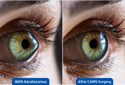
CAIRS (Corneal Allogenic Intrastromal Ring Segments) is an innovative surgical procedure designed to treat keratoconus, a progressive eye disease that causes the cornea to thin and bulge into a cone-like shape. This distortion of the cornea leads to blurry and distorted vision, making everyday activities challenging.
CAIRS involves implanting donor corneal tissue segments into the cornea to provide structural support and improve its shape, thereby enhancing vision and halting the progression of keratoconus. This procedure offers a promising solution for those whose condition has not responded well to other treatments like contact lenses or corneal collagen cross-linking, providing a new lease on life for patients struggling with this debilitating condition. In contrast with traditional corneal transplants, CAIRS reshapes the cornea using ring segments created from donor corneal tissue, offering a more effective and natural correction.
One day, imagine experiencing a clear eyesight that you haven’t had in years. These days, a lot of people with eye diseases like keratoconus or corneal ectasia can actually achieve this because of breakthroughs in eye surgery. A notable example of this is the CAIRS eye surgery. If you or a loved one is thinking about having this operation done, we’ll walk you through everything you need to know, making sure you understand every step and feel confident.
How Does CAIRS Treatment Procedure Work?
With keratoconus, a progressive eye disease, vision becomes distorted as the cornea thins and attains cone-shaped. In order to stabilise and restructure the cornea, corneal ring segments are implanted during the CAIRS operation. The below four points will guide you about thorough rundown of the CAIRS treatment process:
1. Indications
CAIRS is often recommended for patients with:
- Progressive Keratoconus.
- Other corneal ectasias that do not respond well to conservative therapies, such as rigid contact lenses.
- Patients who are not good candidates for corneal collagen cross-linking or other surgical procedures.
2. Preoperative Assessment
Before the surgical treatment, a comprehensive eye examination is performed as follows,
- Corneal Topography is used to map the corneal shape and assess the extent of the ectasia.
- Pachymetry is used to determine corneal thickness.
- Ocular History and Visual Acuity Testing are used to determine the influence on vision and set a baseline.
- The purpose of the contraindications evaluation is to ensure that no conditions, such as active infection or extensive corneal scarring, preclude surgical intervention.
3. CAIRS Procedure
Anaesthesia
- The surgical procedure is usually done under local anaesthesia with topical anesthetic drops.
Creation of Stromal Tunnel
-
A femtosecond laser or a mechanical microkeratome is utilised to create a precise tunnel through the corneal stroma. This tunnel is where the corneal segments will be inserted.
-
The depth and length of the tunnel are carefully estimated using preoperative measurements.
Preparation of Allogenic Segments
- CAIRS corneal segments are created from the donor corneal tissue. These segments are formed into little rings or arcs that offer structural support for the cornea.
- To assure its suitability for implantation, allogenic tissue is treated and sterilised.
Insertion of Segments
- The allogenic corneal ring segments are carefully placed in the stromal tunnel.
- The positioning is critical for producing the desired effect on corneal shape and stability. The severity and asymmetry of the keratoconus determine whether one or two segments are inserted.
Final Adjustments and Healing
- Following insertion, the segments are adjusted to ensure optimum alignment and positioning.
- Antibiotic and anti-inflammatory drops are given to help prevent infection and inflammation.
4. Postoperative Care
- Patients are carefully monitored following surgery, with regular follow-up sessions.
- They are given a regimen of antibiotic and anti-inflammatory eye drops.
- Visual acuity and corneal topography are examined on a regular basis to ensure the procedure’s success and detect any problems at the earliest.
Benefits of CAIRS for Keratoconus
The CAIRS technique has many benefits for individuals with keratoconus, making it a viable choice for managing this degenerative eye disease. Here are the main benefits of CAIRS for keratoconus:
1. Stabilisation of Corneal Shape
- CAIRS gives structural support to the cornea, slowing the course of keratoconus by preventing additional thinning and bulging.
- The use of allogenic segments can result in long-term stabilisation of the corneal shape, eliminating the need for future invasive treatments.
2. Improvement in Vision
- By reshaping and stabilising the cornea, CAIRS can considerably reduce irregular astigmatism, a prominent source of visual distortion in keratoconus patients.
- Many patients report better visual acuity as the corneal shape becomes more regular, resulting in clearer and sharper eyesight.
3. Minimally Invasive Procedure
- CAIRS is less invasive than standard corneal transplantation (penetrating or deep anterior lamellar keratoplasty), which requires more intensive surgery and a longer recovery time.
- It often has a quicker recovery period than more invasive surgical treatments, allowing patients to resume their regular activities sooner.
4. Compatibility with Other Treatments
- CAIRS can be used in conjunction with corneal collagen cross-linking (CXL), which strengthens the corneal collagen fibres. The combination can improve stability and vision.
- It can be adjusted to specific patient demands by adjusting the number and location of the ring segments based on keratoconus severity and asymmetry.
5. Use of Donor Tissue
- The use of allogenic (donor) corneal tissue segments assures greater biocompatibility and lowers the likelihood of adverse responses when compared to synthetic implants.
- Donor tissue blends seamlessly with the patient’s cornea, facilitating natural healing and lowering the risk of rejection or extrusion.
6. Potential for Delay or Avoidance of Transplantation
- By stabilising the cornea early in the disease process, CAIRS can postpone or even eliminate the need for corneal transplantation, a more complicated and hazardous treatment.
- Delaying or postponing transplantation can also save money in the long run, lowering the patient’s overall healthcare burden.
7. Customizability
The technique can be customised to the patient’s individual corneal shape and degree of ectasia. Surgeons can modify the number, size, and positioning of the segments to produce the best results.
Who Is Required to Do This CAIRS Procedure?
The CAIRS procedure should be conducted by a highly skilled eye surgeon with a medical degree and ophthalmology residency. Ideally, the surgeon should have extra fellowship training in cornea and refractive surgery, which allows for specialised competence in treating corneal illnesses and executing advanced corneal procedures. They must be board-certified in ophthalmology and have extensive experience diagnosing and managing keratoconus, as well as knowledge of corneal surgical methods, particularly those using intrastromal implants.
Experience with modern equipment such as femtosecond lasers or mechanical microkeratomes is also required. To guarantee comprehensive patient care, the surgeon should participate in ongoing education to stay up to date on the newest breakthroughs, join relevant professional organisations, and collaborate with a multidisciplinary team of experts. To achieve the best results, effective communication skills are required while meeting with patients, explaining the operation, and giving detailed postoperative care.
Will I See Better After Having CAIRS Surgery?
Many individuals see significant improvements in their vision after CAIRS surgery, while the level of improvement varies depending on the severity of the keratoconus, prior eyesight, and corneal features. It can decrease irregular astigmatism and increase visual acuity, resulting in crisper and sharper vision. Patients frequently report better eyesight, with fewer distortions and glare. The surgeon’s precision in segment placement, adherence to postoperative care recommendations, and corneal health all contribute to the surgery’s outcome. While CAIRS primarily tries to stabilise the cornea and slow disease progression, many patients still require corrective lenses, albeit less strong ones. It is critical to set reasonable expectations and explore possible outcomes with the surgeon.
Is CAIRS the Only Option Left to Improve My Sight, or Are There Other Treatments?
CAIRS is one of various methods for improving eyesight in people with keratoconus and other corneal ectatic conditions. Other eye treatments include glasses and contact lenses, which can correct vision in the early stages; rigid gas permeable (RGP) and scleral lenses, which provide a more consistent refractive surface for moderate to advanced keratoconus; and corneal collagen cross-linking (CXL), which strengthens the corneal collagen fibres and slows disease progression. Furthermore, Intacs (intrastromal corneal ring segments) are synthetic implants used to reshape and stabilise the cornea, similar to CAIRS but with plastic segments instead of donated tissue. The severity of the condition, corneal characteristics, and unique patient demands all influence treatment decisions, which frequently necessitate a consultation with a trained ophthalmologist to establish the best strategy.
How Much Does CAIRS Surgery Cost?
In India, the cost of CAIRS varies based on the patient’s eye features and the type of corneal problem being treated. The severity of keratoconus, as well as the particular corneal shape and thickness, can all have an impact on the procedure’s difficulty and cost. Furthermore, the geographic location, surgeon expertise, and kind of medical facility all have an important impact in deciding the final keratoconus surgery cost. A full consultation with an experienced ophthalmologist is required to provide an accurate cost estimate based on your specific needs and corneal health.
Who Developed the CAIRS Procedure?
Dr. Soosan Jacob, a distinguished ophthalmologist and pioneer in corneal and refractive surgery at Dr Agarwals Eye Hospital, created the CAIRS procedure. Dr. Soosan Jacob is well-known for her unique contributions to ophthalmology, which have helped advance numerous surgical approaches for treating difficult corneal problems. Her CAIRS method, which uses allogenic tissue to stabilise and restructure the cornea, is a revolutionary strategy for treating keratoconus and other corneal ectatic problems.
|
Verified by: Dr. T. Senthil Kumar MBBS MS (Ophthal) (Gold Medallist) FICO |
Reference:
- Jacob S, Agarwal A, Awwad ST, Mazzotta C, Parashar P, Jambulingam S. Customised corneal allogenic intrastromal ring segments (CAIRS) for keratoconus with decentered asymmetric cone. Indian Journal of Ophthalmology/Indian Journal of Ophthalmology. https://pubmed.ncbi.nlm.nih.gov/37991313/
Frequently Asked Questions (FAQs) about CAIRS Eye Surgery
Is CAIRS a new procedure?
Yes, CAIRS is a relatively new treatment for treating keratoconus and other corneal ectatic conditions. Donor corneal tissue rings are implanted into the corneal stroma to offer structural support and improve corneal stability.
Is CAIRS right for everyone with Keratoconus?
CAIRS is not appropriate for everyone with keratoconus. The technique is usually indicated for individuals with progressive keratoconus who have not responded adequately to conservative therapies such as contact lenses. A comprehensive evaluation by a corneal specialist is required to evaluate whether CAIRS is the best option based on the individual’s corneal thickness, shape, and overall eye health
What are the long-term effects of CAIRS?
The long-term effects of CAIRS are still being explored, but initial results indicate that the surgery can give consistent, long-term improvements in corneal shape and vision. The majority of individuals had keratoconus progression halted, and their visual acuity has improved. Ongoing monitoring is required to ensure the stability and health of the cornea.
How does CAIRS compare to other Keratoconus treatments like contact lenses?
CAIRS is a structural solution that strengthens and shapes the cornea, whereas contact lenses, particularly Rigid Gas Permeable (RGP) and scleral lenses, correct vision by providing a smooth refractive surface. CAIRS can reduce or improve the comfort and effectiveness of contact lenses, but they may not completely eliminate the need for corrective lenses.
What are the risks associated with CAIRS?
CAIRS risks include infection, inflammation, segment displacement or extrusion, and the need for additional surgical intervention if issues emerge. As with any surgical operation, there are inherent risks that must be discussed with the surgeon beforehand.
Is CAIRS right for me if I already had corneal collagen cross-linking?
CAIRS (Corneal Allogenic Intrastromal Ring Segments)may be an option for patients who have already undergone corneal collagen cross-linking. The two operations can work together, with cross-linking stabilising the cornea at a biochemical level and CAIRS giving mechanical support while improving corneal shape. A corneal specialist can determine whether CAIRS is appropriate in your unique instance.
How does CAIRS surgery affect night vision?
Night vision may be affected temporarily following CAIRS surgery due to postoperative healing and probable corneal shape alterations. Some patients may have glare and halos at first, but these symptoms usually resolve as the cornea recovers. Long-term night vision results are generally positive, particularly when compared to untreated progressive keratoconus.
What is the recovery process like for CAIRS?
The CAIRS recovery procedure consists of multiple stages. Patients may suffer discomfort, redness, and impaired vision at first, but these symptoms usually subside after a few days. Eye drops are used to prevent infections and relieve irritation. Regular follow-up visits are essential for monitoring healing and the position of the ring segments. Most patients can return to normal activities within a week, although the final visual outcome may take several months as the cornea stabilises and adjusts.

